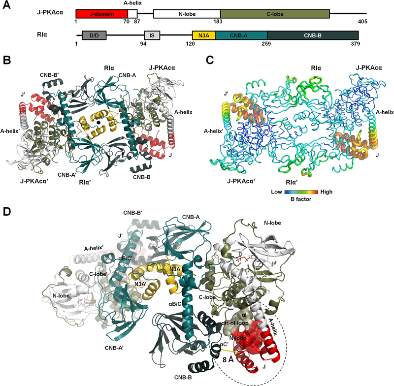
Figure 1. Overall structure of the chimeric RIα2:J-PKAcα2 holoenzyme.
(A) Domain organization and color coding of J-PKAcα and RIα subunits.
(B) Structure of the holoenzyme. One heterodimer is labeled as RIα:J-PKAcα and its two-fold symmetry mate is labeled as RIα’:J- PKAcα’. The two-fold axis position is shown as a solid black circle.
(C) B factor analysis of the holoenzyme.
(D) The J-domain is in close proximity to the CNB-B domain of RIα.

