
Figure 1.
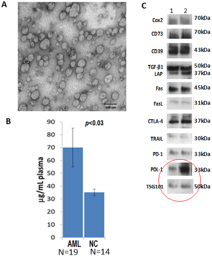
Characteristics of blast-derived exosomes isolated from plasma of patients with acute myeloid leukemia (AML). In A, transmission electron microscopy (TME) image of exosomes isolated by size exclusion chromatography (SEC) from plasma of an AML patient at diagnosis. Note the size heterogeneity of these exosomes. In B, protein levels (calculated per 1mL of plasma) of exosome fractions isolated by SEC from plasma of AML patients at the time of diagnosis and from normal controls. In C, representative Western blots of exosomes from plasma of two AML patients showing the presence of several immunoinhibitory proteins. The red circle calls attention to the presence of PD-L1 in the exosome cargo and of TSG101, an endocytic marker.