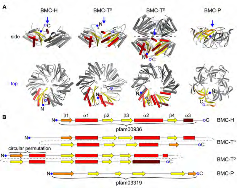
Fig. 1. Component structures of the HO BMC shell.
(A) Cartoon representation of structures of the BMC shell proteins found in the HO shell, viewed from the side (top row) and from the top onto the concave side (bottom row; view point indicated by a blue arrow in the side view). One protein chain each is colored according to secondary structure (helix: red, sheet: yellow, first and last secondary structure elements colored darker) and marked N- and C-termini.
(B) Secondary structure diagrams of the shell proteins depicted in a. BMC-H consists of a single pfam00936 domain. HO-BMC-TS1 is a member of the BMC-TS-type that comprises a fusion of two pfam00936 domains. HO-BMC-TD2 and HO-BMC-TD3 are BMC-TD-type shell proteins that are a fusion of a permuted pfam00936 domain where β4 and α3 are located at the N-terminus. BMC-P contains the all-β-sheet pfam03319 domain.

