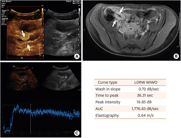
Fig. 1. A case of acute inflammation in a child with Crohn's disease. (A) Contrast-enhanced ultrasound demonstrates avid contrast enhancement in the narrowed terminal ileum. (B) Transverse contrast magnetic resonance imaging of the abdomen demonstrating bowel wall thickening and inflammation in the terminal ileum. (C) Contrast enhancement curve and quantitative parameters generated from the time-intensity curves based on the drawn region of interest in the terminal ileum.
LDRW: local density random walk, WIWO: wash-in, wash-out, AUC: area under the curve.

