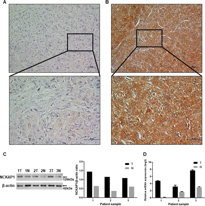
Fig. 1. Immunohistochemistry (IHC), western blotting (WB), and quantitative real-time PCR (qPCR) characteristics of NCKAP1 in hepatocellular carcinoma (HCC) specimens. NCKAP1 was primarily expressed in the tumor cell cytoplasm.
a Representative images of negative NCKAP1 expression in the tumor cytoplasm (×200). The lower panel shows an enlargement of the indicated area (×400). b Representative staining of negative NCKAP1 expression in the tumor cell cytoplasm (×200). The lower panel shows an enlargement of the indicated area (×400). c WB results show that NCKAP1 exhibited higher expression in tumor tissues compared to adjacent normal tissue in patient samples. d qPCR confirms the higher expression of NCKAP1 in tumor tissues

