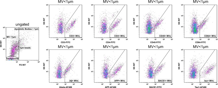
Figure 1.
Gating of microvesicles. The respective antibody was added to CSF, incubated and measured directly and undiluted with flow cytometry. Microvesicles were defined by a gate that excluded background and particles with a higher forward scatter than 1 µm beads. The expression of CD3, CD4, CD45, CD64, Aβ, APP, BACE1, and tau was analyzed within the microvesicle gate (MV < 1 µm). Gates defining the respective antigen positive fraction were drawn in respect to an obviously antigen negative fraction within the same measurement.

