
Figure 2. Variant map of variants in MT-ATP6 mapped by protein domain.
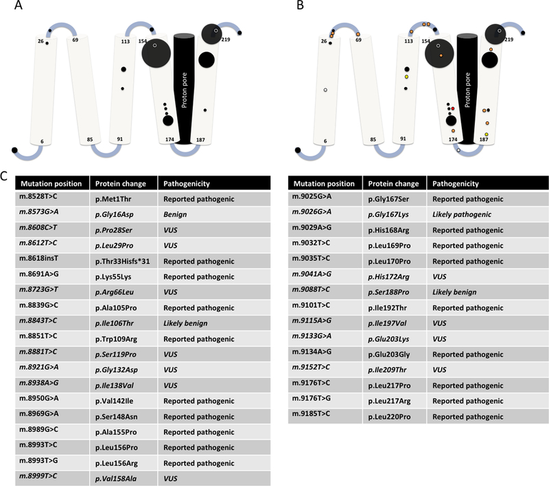
A. Reported pathogenic variants in MT-ATP6 mapped across its five transmembrane domains. The area of each circle is proportional to the number of reported patients. B. Reported MT-ATP6 variants in a new cohort reported in this publication (n=16). White circles represent variants re-classified as benign, yellow circles represent variants re-classified as likely benign, orange circles represent VUS, and red circles represent variants re-classified as likely pathogenic. C: List of all mutations including protein changes and classification.