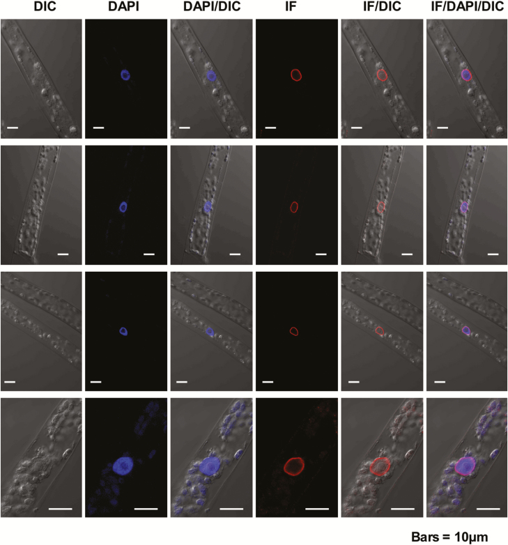
Fig. 5.
Localization of Physcomitrella patens NMCP in protonema cells. Confocal sections of four representative protonemata after staining with the anti-PpaNMCP1 antibody, showing the distribution of PpaNMCP1, chromatin staining with DAPI, and the corresponding differential interference contrast (DIC) images. Overlays of the PpaNMCP1 and DAPI stainings and the corresponding DIC images are also shown. PpaNMCP1 localizes at the nuclear periphery wrapping chromatin. Bars=10 µm. (This figure is available in colour at JXB online.)

