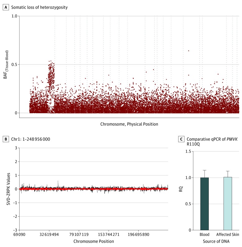
Figure 3. Copy Neutral Loss of Heterozygosity Is Evident in Affected Tissue From Participant 1.
Abbreviations: BAF, B-allele frequency; RQ, relative quantification; SVD, singular value decomposition; ZRPK, z-scores of reads per kilobase. A, B-allele frequency differences between the affected tissue and blood are plotted across the genome for participant 1 by chromosome and physical position. Dashed vertical lines separate individual chromosomes. Somatic loss of heterozygosity on chromosome 1q of participant 1 extends from 14.66 megabases to the telomere, and contains the PMVK gene. B, The CoNIFER program was used to detect copy number variations on chromosome 1. Singular value decomposition of standardized z-scores of reads per thousand bases, per million reads is plotted against positions on chromosome 1 and shows no evidence for copy number variation. C, This is further corroborated by comparative qPCR assessing the PMVK R110Q location. The fold change of the copy number of genomic PMVK from affected skin is calculated using relative quantification (RQ): RQ, 1.01 (range, 0.91-1.13) for affected skin when the blood of participant 1 was used as the reference sample.

