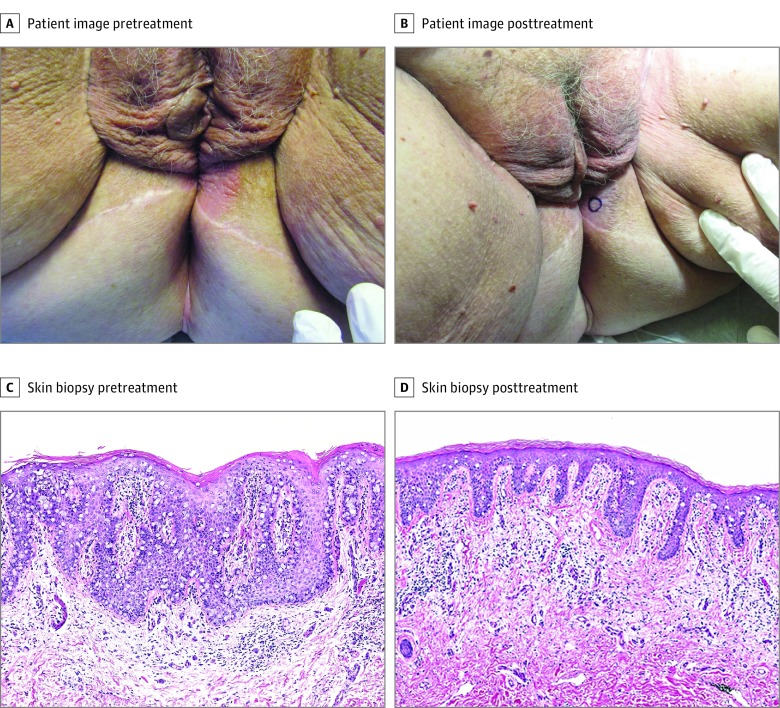
Figure 3. Clinical and Histopathological Images of Refractory Extramammary Paget Disease (EMPD) in Case 2.
A, A 3-cm, well-demarcated, thin erythematous plaque on the left perineum extending to the left labia majora. B, Reduced erythema and peripheral extension of left perineal lesion after 8 months of combination therapy. C and D, Hematoxylin-eosin–stained lesional specimens (original magnification ×100). C, Pretreatment specimen reveals well-developed EMPD with a relatively high density of tumor cells and epidermal hyperplasia. D, After 8 months of combination fluorouracil and calcipotriene treatment, a repeated skin biopsy reveals a lower density of intraepidermal neoplastic cells, a less hyperplastic epidermis, and upper dermal changes that include superficial perivascular edema and lymphocytic inflammation.

