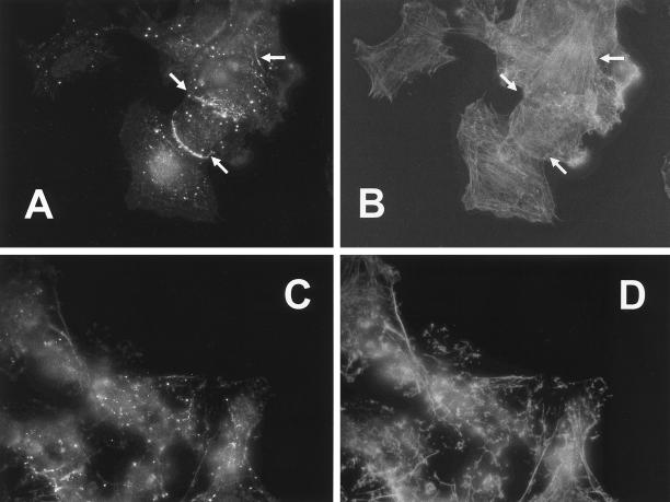
Figure 9.
Disruption of F-actin leads to altered PKCε localization. SK-N-BE(2) cells were treated with 0.6 μM latrunculin B for 20 min whereafter PKCε was visualized with immunofluorescence (A and C) with Alexa Fluor 488-conjugated secondary antibodies, and F-actin was detected with Alexa Fluor 546-conjugated phalloidin (B and D). Cells were analyzed with a fluorescence microscope and images show control cells (A and B) and latrunculin B-treated cells (C and D). Arrows in A and B indicate cortical areas enriched in PKCε. These were invariably found in cortical areas where the cell had contact with another cell.

