Fig. 4.
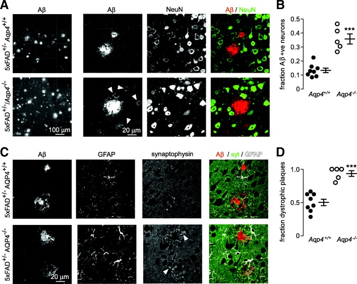
Increased neuronal Aβ uptake and more peri-plaque dystrophic neurites in Aqp4 deficient 5xFAD mice. a Intermediate (left panels) and high (right panels) magnification images showing Aβ uptake in NeuN-labeled neurons surrounding plaques (arrowheads). b The fraction of Aβ-positive neurons surrounding plaques as measured in at least 10 separate plaques from each mouse of each genotype (Aqp4+/+ n = 8, Aqp4−/− n = 5, *** p < 0.001 by unpaired t-test). c Synaptophysin labeled presynaptic dystrophies (arrowheads) surrounding plaques showing increased peri-plaque dystrophies in Aqp4 deficient 5xFAD mice. d The fraction of dystrophic plaques (4 or more large presynaptic dystrophies) determined in 8–10 plaques from each mouse (Aqp4+/+ n = 8, Aqp4−/− n = 5, *** p < 0.001 by unpaired t-test)

