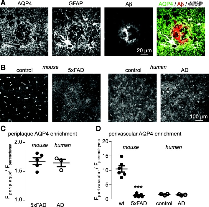
Fig. 5.
AQP4 is increased in peri-plaque astrocyte processes in post-mortem samples from human AD patients. a AQP4, GFAP and Aβ immunofluorescence in inferior frontal gyrus of a human AD patient showing AQP4 enrichment in peri-plaque astrocyte processes. b Lower magnification images showing extensive redistribution of AQP4 away from endfeet in 5xFAD mice compared to age matched wild-type mice, and corresponding images from aged control and AD human patient samples, demonstrating that AQP4 is mostly parenchymal in both cases. c Average enrichment of AQP4 in peri-plaque astrocyte processes, as compared to non-plaque parenchymal areas, in mouse and human AD. d Average enrichment of AQP4 in perivascular regions, compared to the parenchyma, in mouse and human control and AD. Samples from 6 wild-type and 5 5xFAD mice were compared along with 3 control samples and 3 AD human samples. (*** p < 0.001 by unpaired t-test)

