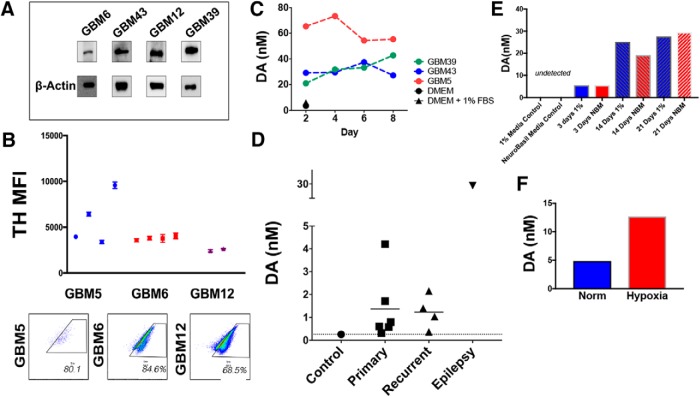
Figure 4.
GBM cells synthesize and secrete dopamine. A, Western blot analysis of TH expression, the rate-limiting enzyme in dopamine synthesis. β-Actin was used as a loading control. B, PDX cells were implanted intracranially into athymic nude mice. Upon sickness from tumor burden, mice were killed and whole brains extracted. FACS analysis of HLA+ tumor cells demonstrates that GBM tumors continue to express TH in vivo. Each dot represents individual mice; FACS analyses were run in technical triplicates. Inset, Scatter plots are representative for FACS results in each cell line tested. C, Conditioned media from PDX GBM cells growing in purified monocultures were analyzed by HPLC-MS for dopamine. Unconditioned DMEM media with and without 1% FBS was used as a control. D, HPLC-MS was performed on GBM samples from patients undergoing resection. Cortical tissue from an epilepsy surgery was used as the control. E, PDX GBM6 cells were growing in either 1% FBS containing media or neurobasal media (NBM). Conditioned media were collected at various days and dopamine levels were calculated using HPLC. F, PDX GBM43 cells were cultured in the presence of either 20% or 1% oxygen (hypoxia) for 3 h. Conditioned media was collected, and dopamine levels determined. Dots represent values from each time point and/or sample tested. Figure 4-1 includes other genes involved in dopamine synthesis (VMAT2, DAT, DDC, TH) measured in patient tumor samples as well as HPLC performed on various PDX subtypes undergoing a TMZ treatment time course.

