Figure 6.
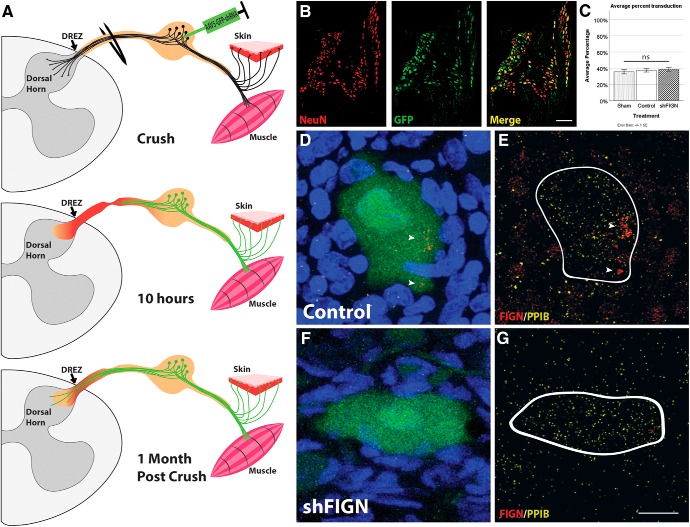
AAV5–U6-shFIGN-CMV-GFP transduces neurons and knocks down fidgetin in vivo. A, A schematic of the in vivo dorsal root crush injury assay. Adult rats have their right dorsal roots crushed from C5 to T1, resulting in complete denervation of the front right paw as a result of the primary injury. The DRGs are injected with AAV5 using a glass micropipette. Hours later, neurons are transduced and the secondary injury begins. Animals are killed 1 month later, by which time DRG neurons are strongly expressing GFP, which is present in both their cell bodies and axons, allowing measurement of regeneration of GFP+ axons beyond the DREZ and into the dorsal horn. B, DRG neurons were identified using a NeuN-antibody, and transduced cells were identified using an antibody to GFP. The merged image exhibits strong GFP fluorescence mainly in neurons. C, Approximately 40% of the neurons were GFP+ across all experimental groups with no significant (ns) differences. D, A GFP+ DRG neuron is surrounded by DAPI+ cells. E, Using RNA Scope, the mRNA of fidgetin (red; white arrows) and PPIB (yellow; positive control) was revealed within the cytoplasm of neurons. Fidgetin mRNA was also located in the nucleus of surrounding non-neuronal cells. F, Neurons injected with AAV5-shFIGN also express the GFP reporter. G, Fidgetin mRNA is knocked down from both the cytoplasm of neurons and nuclei of surrounding non-neuronal cells. Scale bar, 20 μm.

