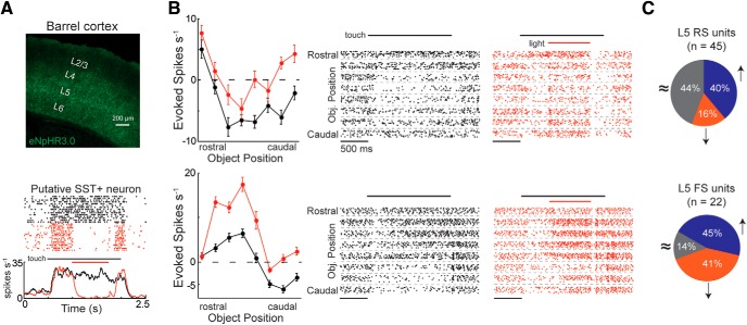
Figure 5.
SST+ deactivation facilitates touch responses in L5 RS units. A, Top, Histological transverse section from an SST-cre mouse expressing eNpHR3.0 tagged with a green fluorophore (YFP). Bottom, Putative SST+ neuron from this mouse that had its activity nearly abolished during the light period. B, Tuning curves and raster plots from two example L5 RS neurons that significantly increase their firing rate during SST+ deactivation (two-way ANOVA, α = 0.05). In the raster plots, only the first 10 trials are shown for each condition to simplify visualization. C, The fraction of L5 neurons that significantly go up (blue), go down (orange), or do not change (gray) during SST+ deactivation (two-way ANOVA, α = 0.05). Error bars are the standard error of the mean.

