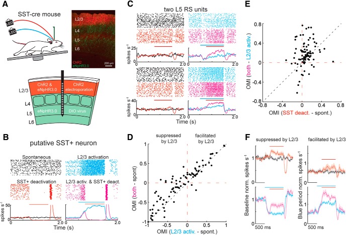
Figure 6.
L2/3's functional impact onto L5 is partially mediated by SST+ interneurons. A, A histological transverse section through the barrel cortex showing ChR2 expression in tdTomato and eNpHR3.0 expression in YFP. DIO-eNpHR3.0 was virally expressed in all layers, whereas ChR2 was expressed only in L2/3 using in utero electroporation. B, Spike raster of a putative SST+ L5 interneuron during spontaneous periods (black), during L2/3 photoactivation (blue), during SST+ photodeactivation (red), and during both presented simultaneously (magenta). C, As in B, but for two example L5 RS units where optogenetic deactivation of SST+ neurons removed L2/3-mediated suppression (top) or enhanced L2/3-mediated facilitation (bottom). D, OMI illustrating the effect of L2/3 activation on spontaneous activity versus the effect of L2/3 activation combined with SST+ deactivation on spontaneous activity. E, OMI scatter plot illustrating the greater disinhibitory effect of SST+ deactivation, whereas L2/3 is photoactivated (p < 0.001, n = 91, Wilcoxon signed-rank test). F, peristimulus time histograms of mean ± SEM of L5 RS unit firing rates during spontaneous activity and the three forms of optogenetic stimulation. Neurons were grouped by the sign of the effect of L2/3 activation and normalized as indicated. Error bars are the standard error of the mean.

