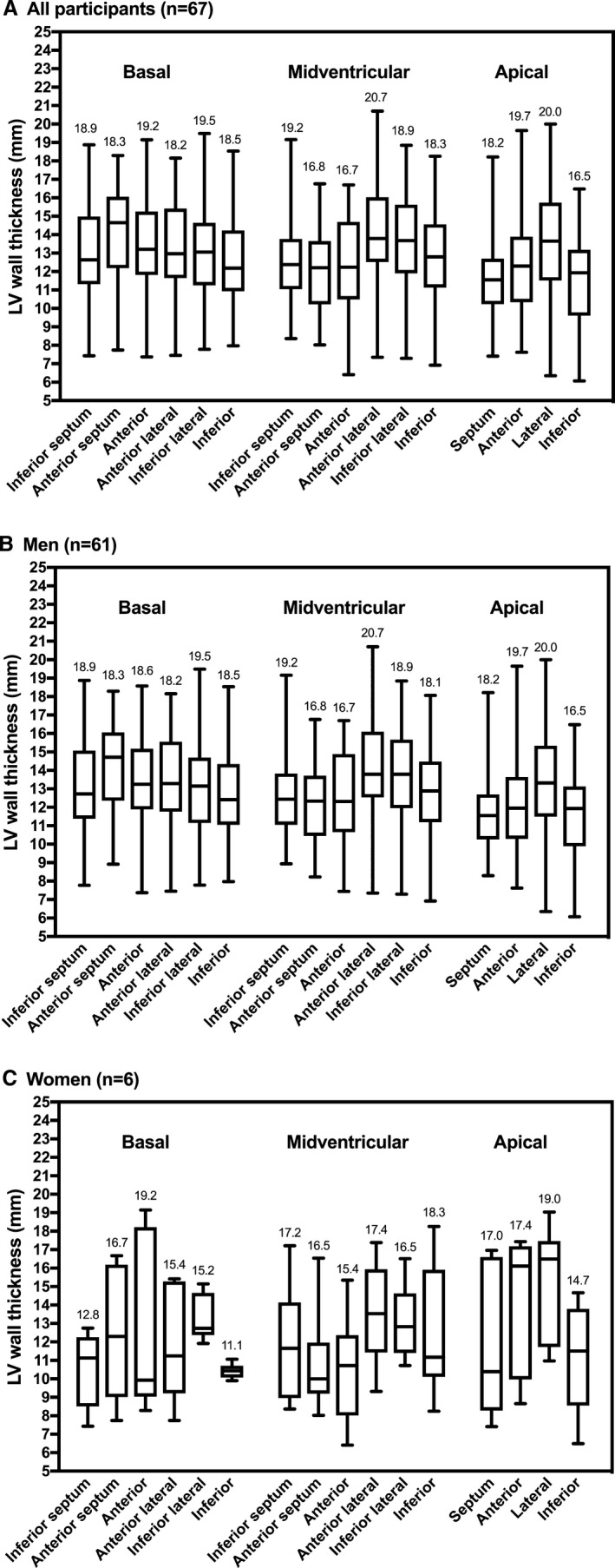
Figure 3.

Distribution of left ventricular wall thickness by segment among all (A), male (B), and female (C) participants with unexplained LV hypertrophy. Maximum wall thickness in millimeters is noted for each segment. LV indicates left ventricular.
