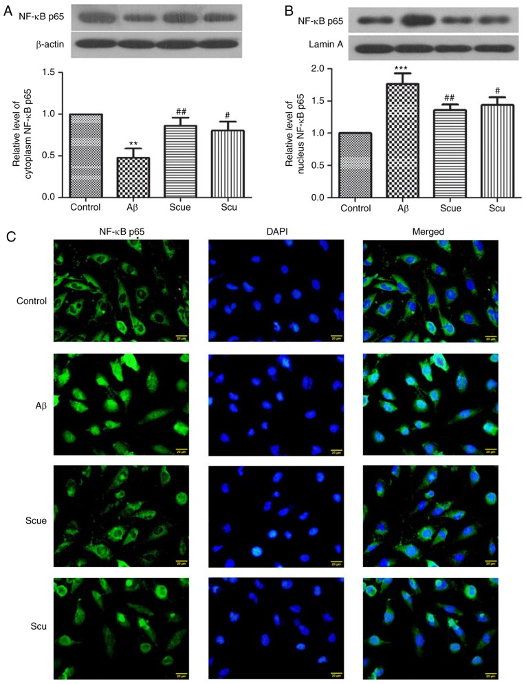
Figure 3.
Scue inhibits the NF-κB signaling pathway. Cytoplasmic expression of (A) NF-κB p65 and (B) nuclear NF-κB p65. Gray scale analyses were conducted using either (A) β-actin or (B) lamin A as an internal reference. Data are presented as the mean ± standard deviation. n=6. (C) Distribution of NF-κB p65 in the cytoplasm and the nucleus were detected by immunofluorescence. NF-κB p65 expression appeared green under a fluorescent microscope and the nucleus was stained blue. Scale bar, 20 µm. Images are representative of repeated experiments. **P<0.01, ***P<0.001 vs. the control group; #P<0.05, ##P<0.01 vs. the Aβ group. NF-κB p65, nuclear factor κ-light-chain-enhancer of activated B cells p65; Scu, scutellarin; Scue, scutellarein; Aβ, amyloid β.

