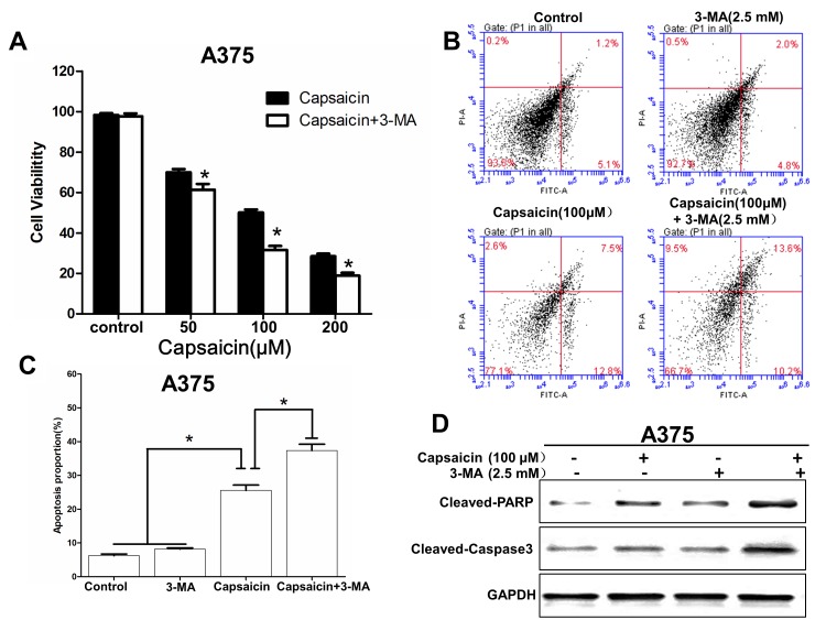
Figure 4.
Inhibition of autophagy increases capsaicin-induced apoptosis. (A) A375 cells were incubated with 3-MA (2.5 mM) for 2 h, and subsequently treated with various concentrations of capsaicin for 24 h, followed by a Cell Counting kit-8 assay to determine cell viability. (B) Following treatment with capsaicin (200 µM) and 3-MA (2.5 mM) for 24 h, A375 cells were incubated with FITC-conjugated Annexin V/PI and the differences in induction of apoptosis were analyzed by flow cytometry. (C) Histograms indicating the proportions of apoptotic cells from three separate experiments. (D) Western blot analysis of the expression levels of the apoptosis-associated proteins cleaved PARP and caspase-3. *P<0.05, compared with capsaicin treatment group or control group.

