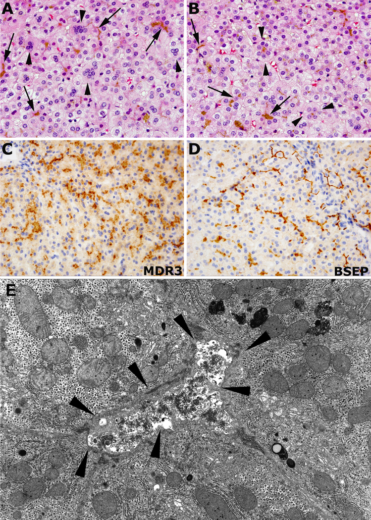
Figure 1.
Representative images of core needle liver biopsies stained with hematoxylin and eosin, and immunohistochemistry performed at our institution. Diffuse hepatocellular damage with loss of the radial orientation of hepatocellular trabecules, multinucleated giant cells (arrowheads in A) and pseudoacinar transformation of hepatocytes (between arrowheads in B), and cytoplasmic and canalicular cholestasis (arrows in A and B). MDR3 (C) and BSEP (D) immunohistochemistry showing thickened and granular reactivity along the canaliculi (in brown color). Representative image of the ultrastructure of the core needle liver biopsy performed at outside institution showing a distended canaliculus (between arrowheads), with luminal osmophilic granular bile and microvillous effacement (E). Abbreviations: BSEP – bile salt export pump.

