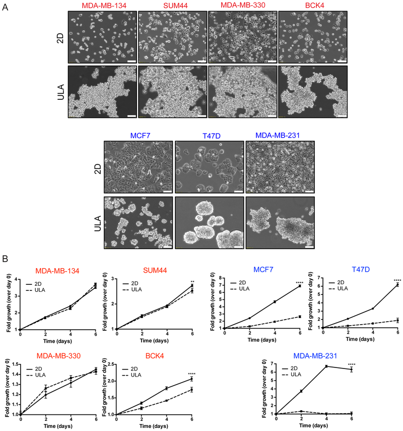
Figure 2. Human ILC cell lines exhibit superior growth in ULA culture than human IDC cell lines.
A. Phase contrast light microscopy images of ILC (red; top) and IDC (blue; bottom) cell lines in 6-well 2D and ULA plates 4 days post plating. Scale bar: 100 μm. B. Relative growth curves showing fold growth normalized to day 0 at each time point over 6 days for ILC (red; left) and IDC (blue; right). Graphs show representative data from three experiments (n=6). p-values are two-way ANOVA comparison of 2D and ULA. * p ≤ 0.05; **** p ≤ 0.0001.

