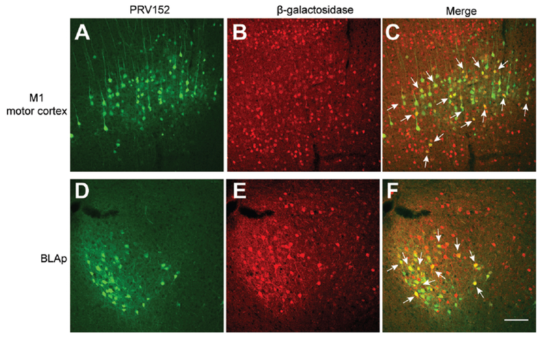
Figure 5. Some BDNF-expressing neurons were polysynaptically linked to iWAT.

(A-C) Representative images showing infection of BDNF neurons in the M1 motor cortex of male BdnfLacZ/+ mice by GFP-expressing PRV152 injected into the iWAT. M1, primary motor cortex.
(D-F) Representative images showing infection of BDNF neurons in the BLAp of male BdnfLacZ/+ mice by GFP-expressing PRV152 injected into the iWAT. BLAp, posterior part of basolateral amygdala.
Immunohistochemistry against β-galactosidase revealed BDNF-expressing neurons in BdnfLacZ/+ mice. Arrows denote some neurons containing both PRV152 and β-galactosidase. Scale bar, 100 μm.
