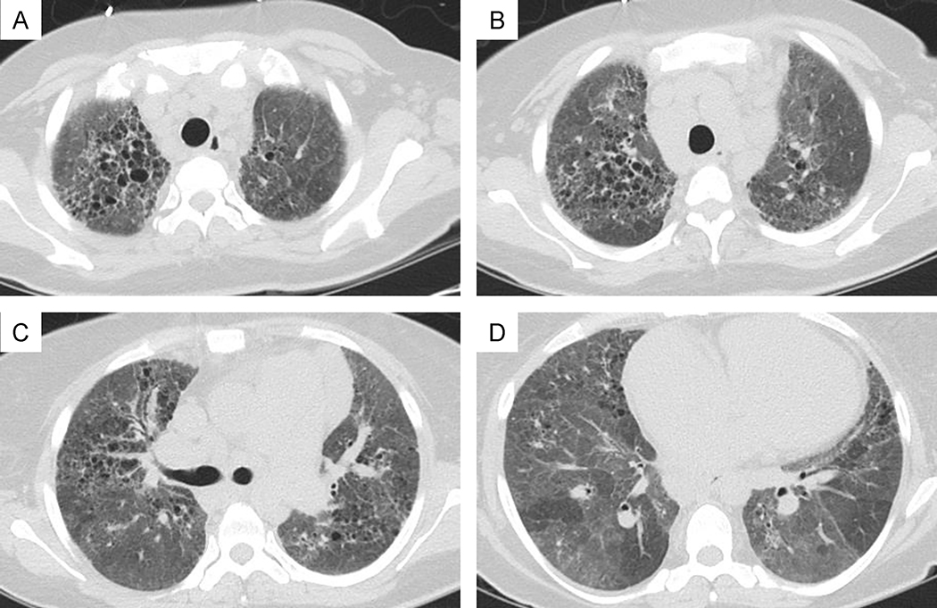
Figure 3: A-D:
High resolution CT of the chest in the same patient shows diffuse upper greater than lower lobe peribronchovascular fine and course reticulation, architectural and pleural parenchymal distortion, traction bronchiectasis and bronchiolectasis, course diffuse ground glass opacification with lobular air-trapping.

