Figure 3.
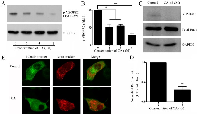
CA inhibits VEGFR2-associated signaling pathway and disrupts cytoskeleton rearrangement. (A) A549 cells were treated with DMSO (control) or indicated concentration of CA for 24 h, prior to assessing VEGFR2 phosphorylation level (Tyr1059) by western blotting. (B) Quantification of WB bands shown in (A). (C) A549 cells were treated with control or indicated concentration of CA for 24 h, prior to assessing Rac1-GTPase by glutathione-S-transferase pull-down and subsequent western blotting. (D) Quantification of phosphor-VEGFR2 level (Tyr 1059) shown in (C), normalized by total VEGFR2. n=4. **P<0.01 and ***P<0.001compared with the 0 µM group. (E) A549 cell were treated with DMSO or CA for 24 h and live cells were stained with Tubulin Tracker™ and Mitotracker® for 5 min. Cells were immediately observed by confocal microscopy (magnification, ×60; scale bar, 10 µm). CA, corosolic acid; Rac1, Ras-related C3 botulinum toxin substrate 1; VEGRF2, vascular endothelial growth factor receptor 2; p, phosphorylated.

