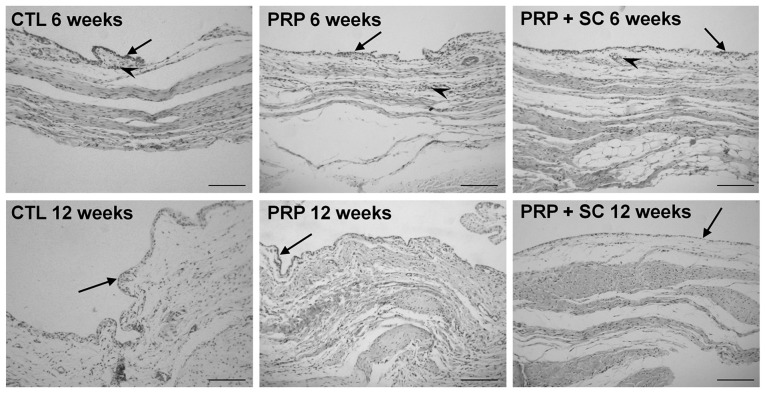
Figure 3.
Representative view of synovial membrane histology from experimental groups stained using H&E. Arrows indicate the layer of synovial lining cells with some stratification in all groups at 6 weeks post-surgery. Inflammatory infiltrate was minimal and was only observed surrounding the blood vessels at 6 weeks post-surgery (arrow heads). Scale bars=100 µm. CTL, control group without treatment; PRP, group treated with platelet-rich plasma; PRP+SC, group treated with PRP plus human dental pulp stem cells.

