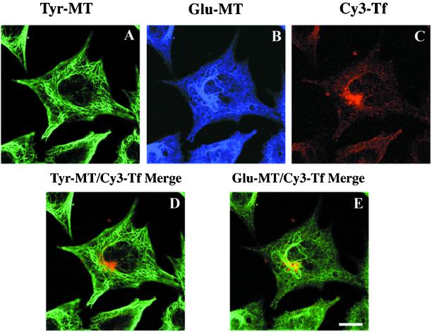
Figure 4.
Confocal microscopy analysis of B104-5 cells triple labeled with Cy3-Tf, anti-Tyr, and anti-Glu tubulin antibodies. B104-5 cells were shifted to 39°C for 4 h before incubation with Cy3-Tf for an additional hour at 39°C. Cells were then fixed, permeabilized, and stained with rat monoclonal anti-Tyr tubulin and rabbit polyclonal anti-Glu tubulin antibodies followed with Alexa488-conjugated goat anti-rat and Cy5-conjugated goat anti-rabbit secondary antibodies. Confocal images of triple-labeled cells were taken as z-series. In A–C, confocal images of Tyr MTs, Glu MTs, and Tf at a single focal plane. In bottom panel, confocal images from the same focal plane, shown in the top panel, were superimposed. For a direct comparison, Cy3-Tf (red) was superimposed either with Tyr-MT (green) (D) or Glu-MT (also green, pseudocolored from Cy5 labeling shown in the top panel) (E). Bar, 10 μm.

