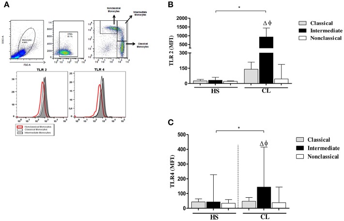
Figure 4.
TLR2 and TLR4 expression in monocytes subsets from CL patients after infection by L. braziliensis. PBMC-derived monocytes from CL patients (n = 08) and HS (n = 08) were infected for 4 h with L.braziliensis (ratio 5:1) stained with CFSE for 4 h. (A) Representative strategy analysis for the selection of monocytes subsets and TLR2 and TLR4 expression. (B) TLR2 expression in classical (CD14highCD16−), intermediate (CD14highCD16+), and non-classical monocytes (CD14lowCD16+) from CL patients and HS after infection by L. braziliensis.(C) TLR4 expression in monocytes subsets from CL patients and HS after infection by L. braziliensis. Data are represented by the median of the mean intensity of fluorescence (MIF). (Δ) Intermediate vs. classical monocytes, P < 0.01 (φ) Intermediate vs. non-classical monocytes, P < 0.01. Mann-Whitney and Kruskal-Wallis with Dunn's post-test was used for statistical test was used for statistical analyses (*P < 0.05).

