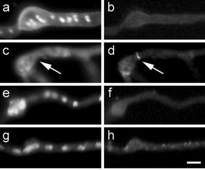
Figure 3.
AspB localization in sep mutants grown at nonpermissive temperature. Chitin and nuclear localization with Calcofluor and Hoechst, respectively (left column). AspB localization (right column). (a and b) sepA. (c and d) sepD. (e and f) sepG. (g and h) sepH. Arrows mark AspB localization in sepD. Bar, 5 μm.

