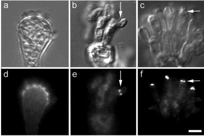
Figure 7.
AspB localization in the conidiophore. DIC images (top row). AspB localization (bottom row). (a and d) AspB localizes at the vesicle/metulae interface as metulae emerge. (b and e) AspB localizes at the metula/phialide interface. Arrow marks emerging phialide. (c and f) AspB localizes to the phialide/conidiospore interface. Arrow marks emerging conidium. Bar, 5 μm.

