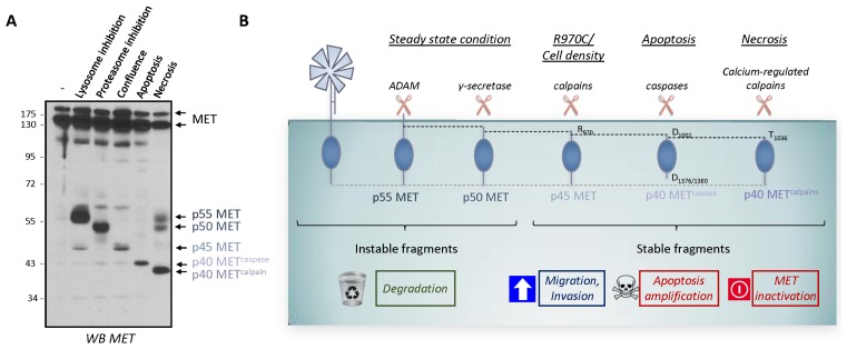
Fig. 2.
Unstable and stable intracellular MET fragments produced by proteolytic cleavages. (A) MCF-10A cells were subjected to the following treatments: incubation with a lysosome inhibitor (5 nM bafilomycin for 5 h); incubation with a proteasome inhibitor (10 μM lactacystin for 5 h); culturing to high density (confluence); incubation with an apoptosis inducer (1 μM staurosporine for 6 h); and induction of necrosis (with 1 μM ionomycin for 1 h). Cell lysates were analyzed by western blotting with an antibody directed against the kinase domain of human MET. Arrows indicate the positions of MET and of the different MET fragments, p55 MET, p50 MET, p45 MET, p40 METcaspase, and p40 METcalpain (see (58) for detailed experimental procedures). (B) Schematic representation of the known unstable and stable intracellular MET fragments generated by proteolytic cleavages.

