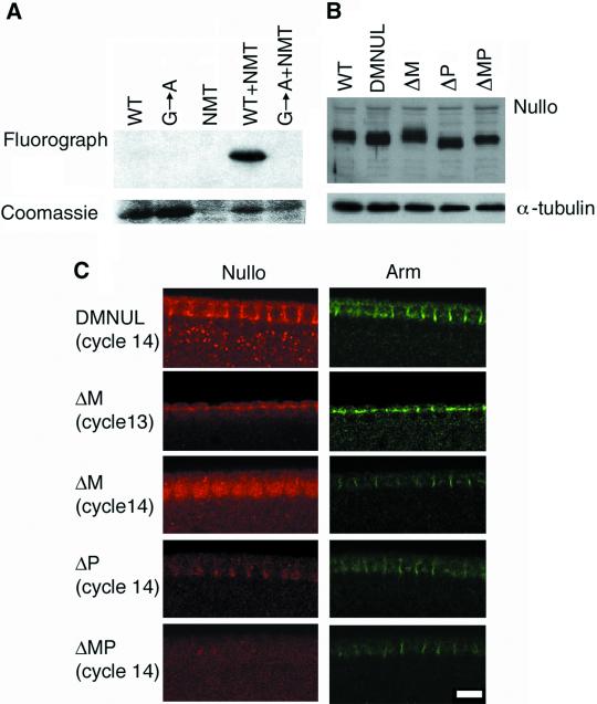
Figure 5.
The N-terminal region is required for Nullo localization. (A) A fluorograph of [3H]myristate–labeled extracts from bacteria expressing Nullo, Nullo containing the G2A mutation, yeast N-terminal myristoyltransferase (NMT), Nullo plus yeast NMT, or Nullo G2A plus NMT. Nullo is labeled with myristate in an NMT-dependent manner, and this labeling is blocked by the G2A mutation. Coomassie-stained Nullo protein is shown as a control. (B) Extracts from embryos expressing only the deletion form of Nullo were immunoblotted using anti-Nullo antibodies. Embryos carrying ΔM, ΔP, and ΔMP show levels of expression equivalent to that of the endogenous Nullo protein and the wild-type nullo transgene (DMNUL). (C) Localization of N-terminal mutants. Embryos lacking endogenous Nullo were stained with anti-Nullo antibodies and counterstained with Arm to visualize the basal junction. The full-length protein produced by the DMNUL transgene localizes to the cellularization front and shows punctate cytoplasmic staining. During cycle 13, ΔM localizes to the membrane of the pseudocleavage furrows and shows weak staining in the nucleus. During cellularization (cycle 14) ΔM relocalizes to the nucleus leaving only weak staining at the cellularization front. ΔP shows a moderate level of staining at the cellularization front but has relatively little punctate cytoplasmic staining. ΔMP has a diffuse cytoplasmic distribution in the cortex of the embryo. Scale bar, 10 μm.

