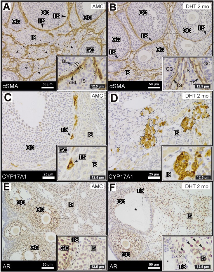
Figure 2.
Ovaries of DHT-treated females exhibit changes in AR localization in the theca and interstitial compartments. (A, B) The vascular cell marker, αSMA (ACTA2) (34) was used to demarcate the external theca because it did not exhibit major changes on DHT treatment (B). (C, D) CYP17A1 was expressed (36) exclusively in specific cells within the theca layer but was not present in granulosa cells or the stromal compartment. After DHT treatment, clusters of CYP17A1+ cells were randomly dispersed in the stroma. (E, F) AR, stained by IHC (35), was nuclear and expressed at high levels in most granulosa cells in control ovaries. AR was nuclear in some theca cells but appeared diffuse and cytoplasmic in the interstitial compartment. On treatment with DHT, AR was nucleated in a subpopulation of interstitial cells (black arrow, inset); an AR− population of cells also expanded (open arrow, inset). N > 3 for all conditions. BL, basal lamina; GC, granulosa cell; IS, interstitial stroma; TE, theca externa; TI, theca interna; TS, theca stroma (TI and TE); αBL, α-smooth muscle actin boundary.

