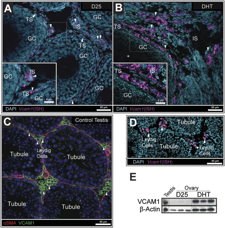
Figure 4.
Vcam1 transcripts were detected in ovarian theca and interstitial cells but not in granulosa cells. (A) ISH in D25 mouse ovaries detected low Vcam1 expression in a few theca cells within the ovary. (B) In contrast, Vcam1 expression was highly elevated in both the theca and stroma of DHT-treated mice. (C) IF images show that VCAM1+ Leydig cells were in contact with VCAM1−/αSMA+ cells lining the seminiferous tubule (31, 34). (D) Vcam1 mRNA was localized exclusively to Leydig cells as determined by ISH. (E) Western analyses of tissue lysates showed that VCAM1 was low in ovaries of D25 control mice but highly induced in ovaries of DHT-treated mice to levels observed in testes from adult mice, as a control. β-Actin was used as a loading control (32). N > 3 for all conditions.

