Figure 8.
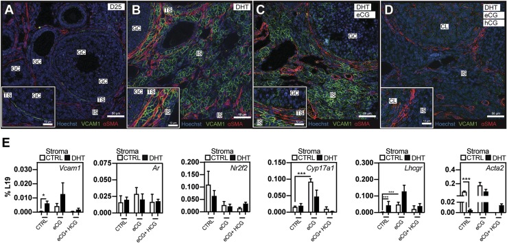
Markers of theca cell function, including VCAM1, are increased by eCG and inhibited by a luteinizing dose of hCG, regardless of DHT treatment. (A) In D25 controls, VCAM1 was expressed (31) at low levels in theca cells of a developing follicle internal to the fibroblastic theca (αSMA+) (34). (B) Ovaries of DHT-treated females exhibited abnormal Vcam1 localization in the theca and interstitial compartment of the ovary. (C) When eCG was administered to DHT-treated females, a small enhancement/compaction of VCAM1+ cells occurred in both the theca and interstitial stroma, whereas (D) a subsequent luteinizing dose of hCG potently abrogated VCAM1 staining in the stromal compartment as well as in theca cells surrounding small growing follicles. (E) To determine the impact of cotreatment with DHT and gonadotropins, and to more precisely document the general physiological timing of VCAM1 expression, qPCR analyses on ovarian RNA prepared from mice on single and combination treatments with DHT and/or eCG and hCG were carried out. Under these conditions, stromal Acta2 expression is inhibited by DHT treatment, but it is reexpressed on treatment with eCG, suggesting a link to stromal remodeling. Whereas Nr2f2 levels decreased in response to eCG/hCG treatments, Ar mRNA remain unchanged. Vcam1, Lhcgr, and Cyp17a1 theca markers were induced on treatment with eCG and conversely suppressed when treated with ovulatory hCG. N = 3 for all conditions. *P < 0.05, ***P < 0.01 by Student t test. GC, granulosa cell; IS, interstitial stroma; TS, theca stroma.

