Figure 5.
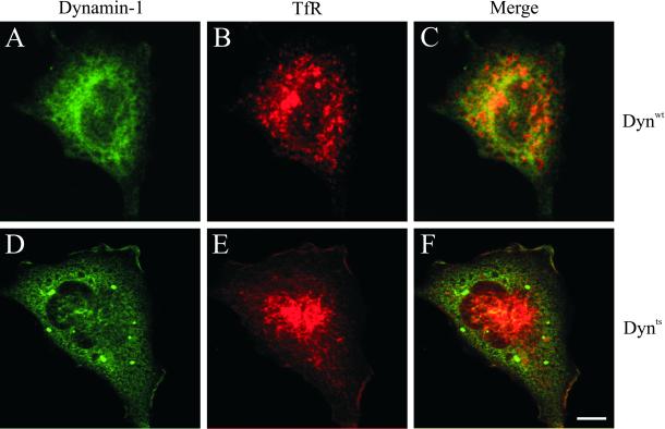
dynts cells redistribute intracellular TfR. dynwt (A–C) and dynts (D–F)-expressing cells were incubated for 30 min at 38°C, fixed, and immuno-double–labeled for dynamin-1 (green) and TfR (red). Integrated views of entire cells were obtained by superimposition of 25 sequential 0.3-μm optical sections. Note that in dynts cells dynamin-1 accumulated at the edge of the cell and in large aggregates. TfR accumulated at the plasma membrane as well as in tubular endosomes in the perinuclear area. The latter structure apparently also accumulated dynts. Bar, 10 μm.

