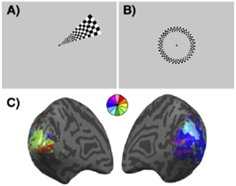
Figure 1:

Retinotopic mapping. (A) Example of a wedge stimulus used to map polar angle visual preferences. Subjects fixate on the central dot while the flickering checkerboard wedge is presented in each of 12 non-overlapping polar angles multiple times. (B) Example of a ring stimulus used to map eccentricity visual preferences. Subjects fixate on the central dot while the flickering checkerboard ring is presented in each of 6 non-overlapping eccentricities multiple times. (C) Example of a retinotopic map from a stroke patient with a visual field cut. The map is pseudo-colored based on each voxel’s preferred wedge location.
