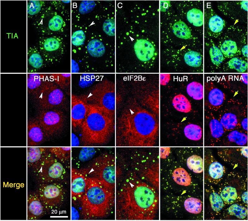
Figure 3.
Other factors present or absent from stress granules. DU145 cells were stimulated with arsenite as described in Figure 2 and processed for immunofluorescence. Each vertical column shows views of the same field. Top row: TIA-1 or TIAR (green). Middle row (red): PHAS-I (eIF4E-binding protein) (A), HSP27 (B), eIF2Bε (C), and HuR (D); poly(A)+ RNA (E). Bottom row, merged views. Yellow arrows point out individual stress granules in which both the red and the green signals overlap; white arrowheads indicate the position of SGs that only contain TIA. Nuclei are stained blue. Bar, 20 μm.

