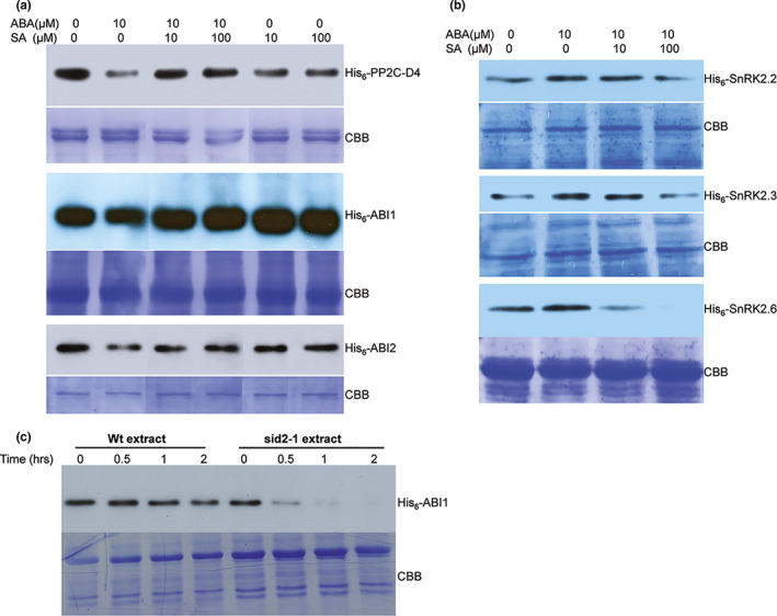
Figure 4.

SA alters ABA‐mediated turnover of PP2Cs and SnRK2s. (a) Cell‐free degradation assay using approximately 100 μg of total protein extracts prepared from 10‐day‐old Arabidopsis seedlings supplemented with 500 ng of His6‐tagged PP2Cs (PP2C‐D4, ABI1, and ABI2) and indicated concentrations of ABA, SA, or ABA+SA. (b) Cell‐free degradation assay using approximately 100 μg of total protein extracts prepared from 10‐day‐old Arabidopsis seedlings supplemented with 500 ng of His6‐Sumo‐tagged SnRK2s (SnRK2.2, 2.3, and 2.6) and indicated concentrations of ABA or ABA+SA. For a and b, the degradation assay was carried out at 30°C for 3 hr. All lanes shown are from the same experiment; some lanes unrelated to this study have been removed and lanes were then merged for clarity of presentation. (c) Cell‐free degradation assay using approximately 100 μg of total protein extracts prepared from 10‐day‐old wild‐type or sid2‐1 Arabidopsis seedlings supplemented with 500 ng of His6‐tagged ABI1. Samples were taken after 0, 0.5, 1, or 2 hr of incubation; proteolysis was stopped by the addition of SDS‐PAGE buffer. Proteins were detected by immunoblotting using an α‐His6‐HRP antibody. Staining with Coomassie Brilliant Blue (CBB) staining of the gel served as a loading control. The experiments were independently repeated at least twice
