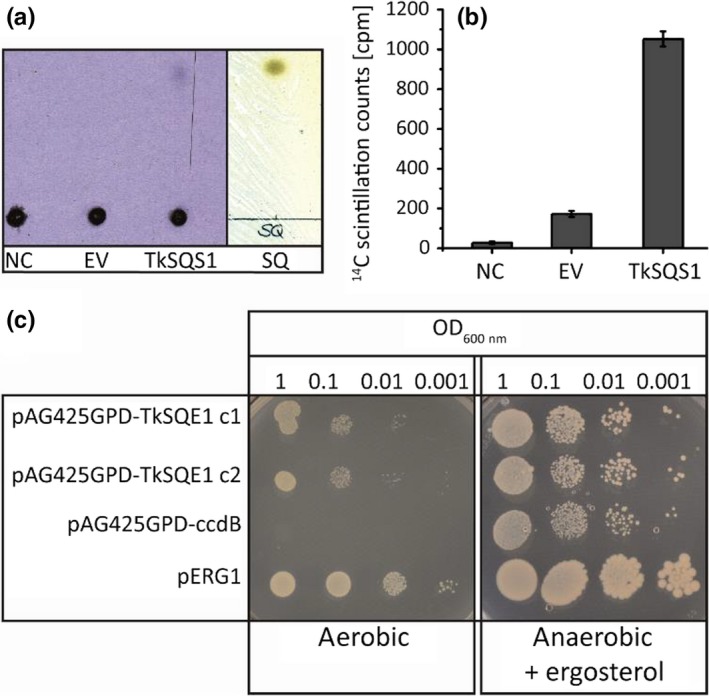
Figure 5.

Functional analysis of TkSQS1 and TkSQE1. TkSQS1 (a–b) activity assay and TkSQE1 complementation (c). (a) Phosphor imaging with negative control (NC), empty vector control (EV) and TkSQS1 protein extracts and corresponding reversed‐phase TLC plate showing squalene standard visualized with iodine vapor. (b) Silica was scraped from the TLC plate at the height of the squalene standard, and radioactivity was measured by scintillation counting. (c) Complementation of the yeast strain KLN1 (Δerg1) to confirm the activity of TkSQE1. The yeast was transformed with the vector pAG425GPD‐TkSQE1 or the negative control vector pAG425GPD‐ccdB. KLN1‐pERG1 complemented with the endogenous ERG1 served as a positive control. Two colonies of KLN1‐TkSQE1 and one colony of KLN1‐pERG1 and KLN1‐ccdB were selected, dropped out, and incubated for 3 days under anaerobic conditions in the presence of ergosterol, or incubated for 5 days under aerobic conditions without supplements
