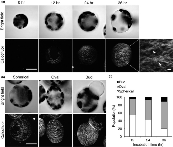
Figure 1.

Cell wall regeneration in protoplasts derived from mesophyll cells of Arabidopsis rosette leaves. (a) Time course of cell wall regeneration in protoplasts. The protoplasts were incubated for 0, 12, 24, or 36 hr and stained with Calcofluor. Stained cells were observed under a bright‐field microscope (top) or a laser scanning confocal microscope (bottom). Inset in the image of protoplasts incubated for 36 hr is magnified on the right; arrowheads indicate bundles of cellulose fibrils. (b, c) Shapes of protoplasts observed under a bright‐field (top) or scanning confocal microscope (bottom) (b), and the population (%) of protoplasts of each shape (c). Bar = 20 μm or 1 μm (magnified image in (a)). n ≥ 126
