Figure 4.

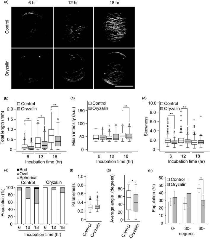
Effects of oryzalin on cell wall regeneration. (a) Representative images of the cellulose network in protoplasts incubated for 6, 12, or 18 hr in the presence or absence of oryzalin. After incubation, the protoplasts were stained with Calcofluor. (b–d) Total length (b), mean intensity (c), and skewness of intensity distribution (d) of the cellulose network regenerated from protoplasts incubated for 6, 12, or 18 hr in the presence or absence of oryzalin. (e) Changes in the populations of protoplasts with three different shapes during incubation for 6, 12, or 18 hr in the presence or absence of oryzalin. (f, g) Parallelness (f) and average angle (g) of the cellulose network with respect to the long axis of oval‐shaped protoplasts incubated for 24 hr in the presence or absence of oryzalin. (h) Effects of oryzalin on the population of oval‐shaped protoplasts with different average angles. Protoplasts were incubated for 24 hr, and oval‐shaped protoplasts were classified according to the average angle (0–29°, 30–59°, 60–90°). Individual populations are shown. Bar = 20 μm. Significance was determined by Mann–Whitney test. **p < .01, *.01 ≤ p < .05. n ≥ 106
