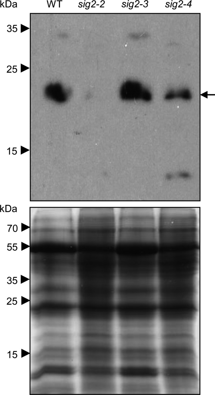
Figure 6.

Accumulation of NDH18 proteins in wild‐type and sig2 mutants. Western blot analysis was performed using anti‐NDH18 antibody (Top panel). Total soluble proteins were extracted from rosette leaves of 40‐day‐old plants grown on soil at 22°C under white light (100 μmol m−2 s−1, long‐day condition with 8 hr dark/16 hr light cycle), and were resolved on 15% SDS‐PAGE gel (Bottom panel). An arrow indicates NDH18 proteins (estimated as 23.8 kDa)
