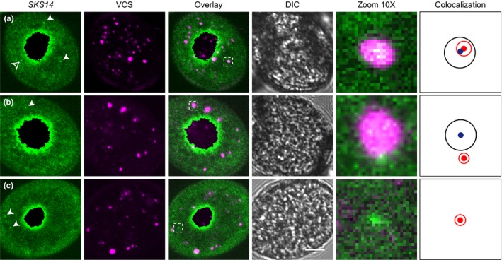
Figure 4.

Colocalization of SKS14 mRNA with RFP‐VCS. (a) Confocal image of a representative mature pollen grain showing high colocalization between SKS14 mRNA and a RFP‐VCS body. (b) Confocal image of a representative mature pollen grain showing a SKS14 mRNA cytoplasmic granule contiguous to a RFP‐VCS body. (c) Confocal image of a representative cytoplasmic granule with no relationship with any RFP‐VCS body. In the left panels, white arrowheads show examples of cytoplasmic granules identified by MATLAB while empty arrowheads show cytoplasmic aggregates not detected by MATLAB. The insets in the merged (“Overlay”) column are enlarged on the 10X panels. In the “Colocalization” column, the blue point and black circle indicate the localization and size of the VCS body, respectively, and the red point and circle correspond to the SKS14 mRNA granule. DIC images are shown. Size bar, 5 μm
