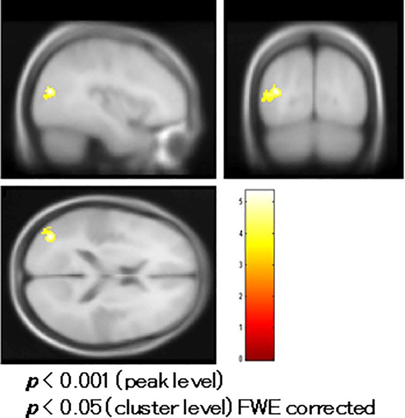
Fig 5. Comparison of functional magnetic resonance imaging (fMRI) between patients and controls.

Patients with schizophrenia had reduced blood oxygenation level-dependent (BOLD) activity in the left middle temporal gyrus (Brodmann area/BA 39) compared to controls in a t-test with age and education as covariates.
