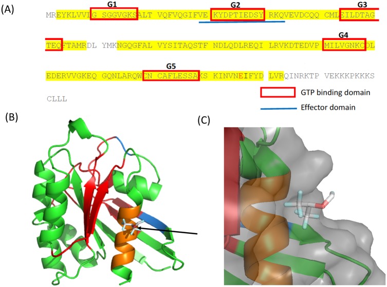
Fig 5. Photolabeling of Rap1 by using azi-sevoflurane.
(A) Photolabeling experiment was performed using azi-sevoflurane. Sequences covered by mass spectrometry is highlighted in yellow. The adducted residue is shown in red. Red box denotes high conserved GTP-binding domain among Ras-related proteins. The blue line denotes effector binding site. (B) Rigid docking of sevoflurane was performed on Rap1. Arrow shows docked sevoflurane. GTP binding domains were indicated in red. The effect binding domain was shown in blue (overlapping G2 domain is shown in red). Residues within 4A from the docked sevoflurane were shown in orange. (C) Sevoflurane binding pocket is shown. The surface is shown in light gray.

