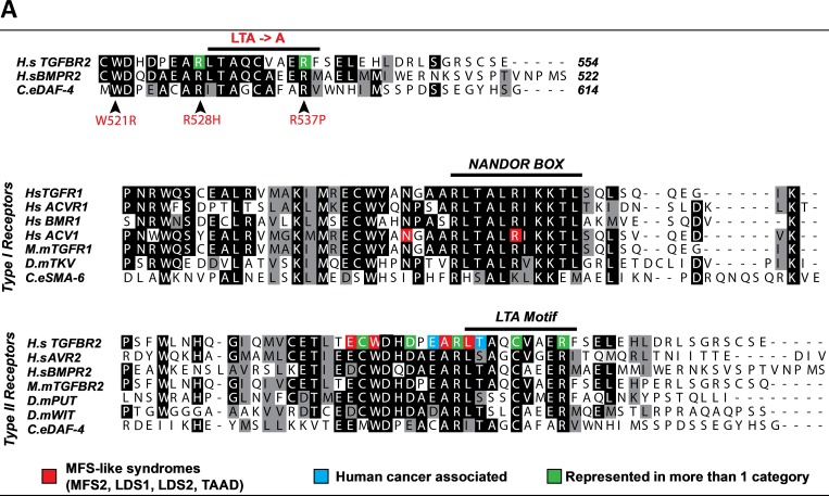
Fig 1.
A. Top Panel: Amino acid alignment of the LTA motifs of the human type II TGFβ and BMP receptors with the type II receptor of C. elegans. Arrows indicate the three mutations we have examined in detail in this study and the region where the LTA motif was substituted with alanines. Middle and Lower Panels: Amino acid alignments of the various type I and type II TGFβ receptors highlight the sequence conservation at the NANDOR box and LTA motif respectively. Black indicates identity, grey indicates conserved changes. For both panels, the various colored boxes identify the residues found mutated in either MFS-like patients, cancers, or in multiple categories (red, blue, orange and green respectively). As observed, all mutations exist as missense mutations in the type II TGFβ receptor.

