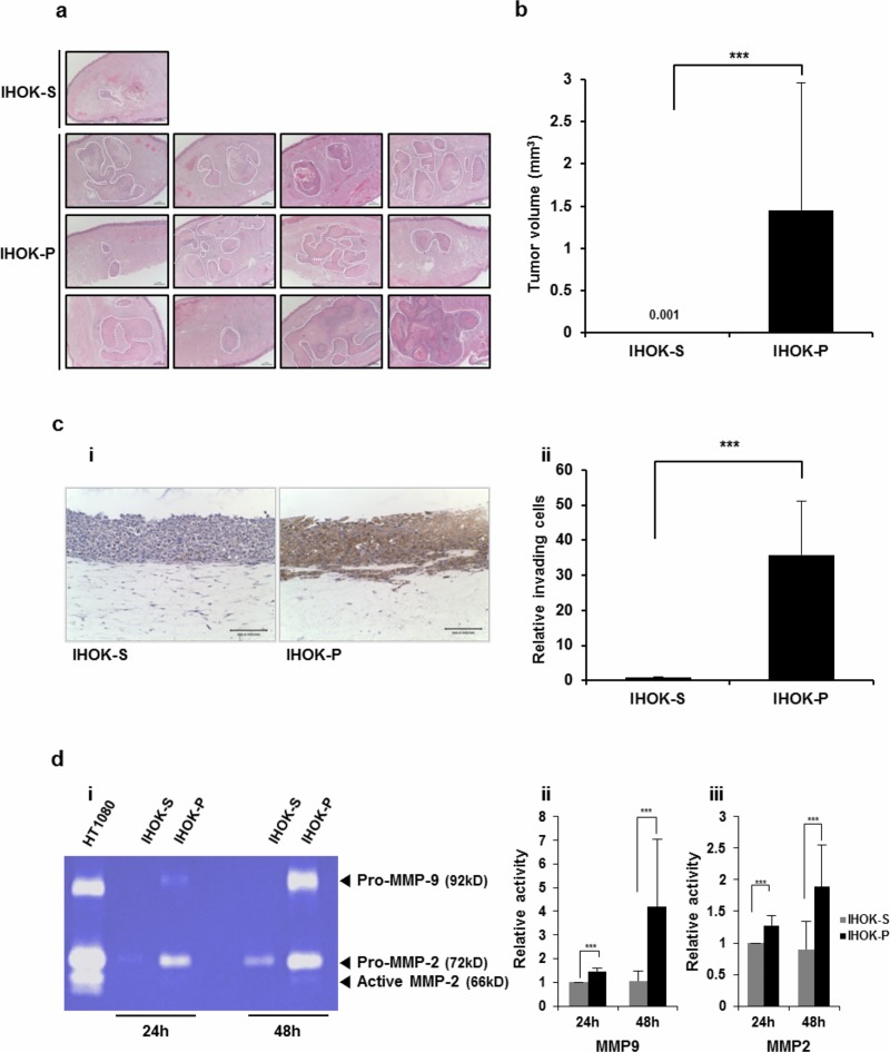
Fig 2. Compared with IHOK-S, IHOK-P had higher tumorigenicity, higher invasiveness, and higher MMP expression.
(a) Histological sections of mice tongues injected with IHOK-S or IHOK-P cells. IHOK-S or IHOK-P cells (5 × 10⁵) were injected into the dorsal tongue of nude mouse (magnification ×40; scale bar, 100 μm). The tumor margin is indicated by a dashed line. (b) Difference in average tumor volume between mice tongues injected with IHOK-S and those injected with IHOK-P. The results were analyzed by the Mann-Whitney U test (***P < 0.001). (c) i) Invasive activity of IHOK-S and IHOK-P cells was determined by 3-dimensional organotypic culture with immunohistochemical staining for cytokeratin (magnification ×200; scale bar, 100 μm). ii) IHOK-P cells more readily invaded into dermal equivalent. All invaded cells were counted by light microscopy. The results were shown as mean ± SD (n = 3) and analyzed by the Mann-Whitney U test (***P < 0.001). Note that this experiment was performed to measure the invasiveness of IHOK-S and IHOK-P, not to measure the level of cytokeratin staining. Cytokeratin was not expressed in IHOK-S, and thus IHOK-S was poorly stained. (d) i) Enzymatic activities of MMP-2 and MMP-9 were measured in IHOK-S and IHOK-P using zymography. IHOK-P cells showed higher MMP-9 (ii) and MMP-2 (iii) activities compared with IHOK-S cells for both 24-hour and 48-hour incubation. HT1080 was used as a positive control. The results were shown as mean ± SD (n = 3) and analyzed by the Mann-Whitney U test (***P < 0.001).

