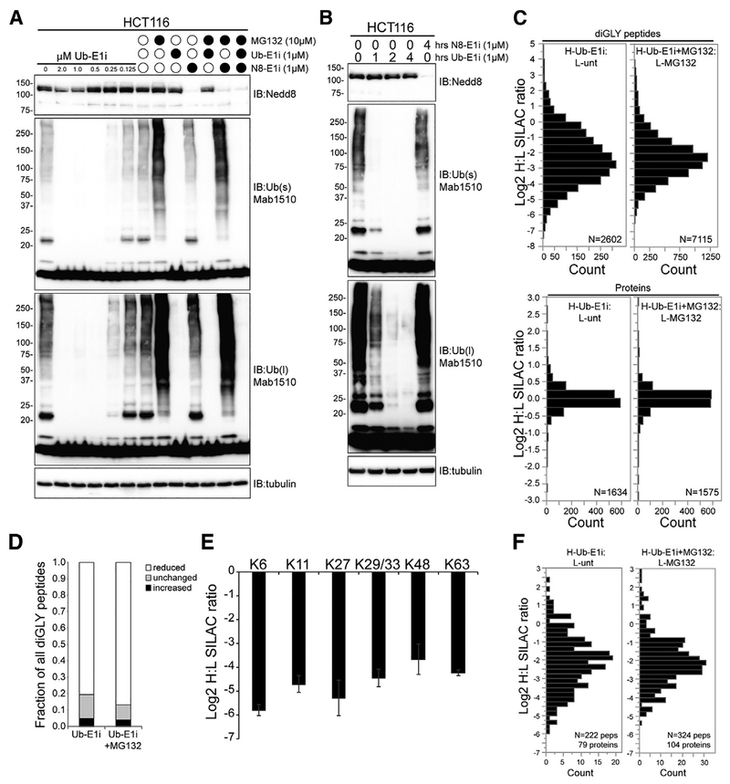
Figure 2. The Ubiquitin-Activating Enzyme Inhibitor TAK-243 Rapidly Ablates Ubiquitin Conjugates in Cells.
(A and B) Immunoblot of Nedd8, ubiquitin, and tubulin in whole-cell extracts from HCT116 cells treated with Ub-E1i or N8-E1i alone (A and B) or in combination with MG132 (A) as indicated. s and l denote short and long exposures, respectively.
(C) Log2 H/L for all diGLY-modified peptides (top) and total proteins (bottom). Heavy-labeled cells were treated with Ub-E1i (1 μM) with or without MG132 (10 μM) for 4 h.
(D) The fraction of all quantified diGLY-modified peptides that increased, decreased, or remained unchanged upon Ub-E1i treatment with or without proteasome inhibition.
(E) Log2 H/L corresponding to diGLY-modified peptides from ubiquitin after Ub-E1i treatment alone. The individual ubiquitin-modified lysine that was quantified is indicated. Error bars denote SEM of multiple quantification events for a given peptide.
(F) Log2 H/L of all diGLY-modified peptides quantified from known SG proteins. Heavy-labeled cells were treated with Ub-E1i (1 μM) with or without MG132 (10 μM) for 4 h.

