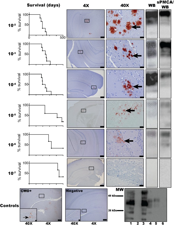
Fig 4. Dilutional mouse bioassay of CWD+ cervid brain into Tg(CerPrP) 5037 mice: survival curves, IHC, WB, and sPMCA/WB prion detection.
PrPSc is shown in the brains of mice inoculated with dilutions 10−2–10−6 of CBP6 (300 μg-30 ng) using IHC (red deposits indicated with arrows), WB, and sPMCA/WB (5th round product) (S1–S3 Figs). PrPSc was not detected in mice inoculated with the 10−7 CBP6 dilution (3 ng) nor in n = 12 negative control mice inoculated with naïve cervid brain. Bottom panels (left to right) show IHC PrPSc deposition in the positive control mouse brain (IC-inoculated 30 μl 10% CWD+ cervid brain homogenate) and no deposition in the negative control mouse (IC-inoculated 30 μl 10% naïve cervid brain homogenate). Control western blot (bottom right) shows complete PK digestion (50 μg/ml) of PrPc in the unamplified (lane 2; without PK in lane 1) and amplified (lane 6) negative control mouse (IC-inoculated 30 μl 10% naïve cervid brain homogenate). PrPSc is revealed after PK digestion (50 μg/ml) in unamplified and sPMCA amplified CWD+ mouse brain (IC-inoculated with 30 μl 10% CWD+ cervid brain homogenate) (lanes 4 and 5; unamplified sample without PK in lane 3). IHC image objectives are 4X (scale bar = 200 μm) and 40X (scale bar = 20 μm).

